


01
The hidden predator.
Screening for melanoma is not screening. It is a hunt for a predator you can’t see. Most melanomas do not look like the pictures you’ve seen. They look like every other spot on the skin.
That isn’t what most people know. The common understanding is melanoma should stand out, and the literature would agree with this: one US study found more than half of melanomas were found by patients themselves,1 and in Australia about two-thirds aren’t picked up at a routine skin check.2
I believe this picture is wrong and may be a large part of why screening hasn’t brought melanoma deaths down.3
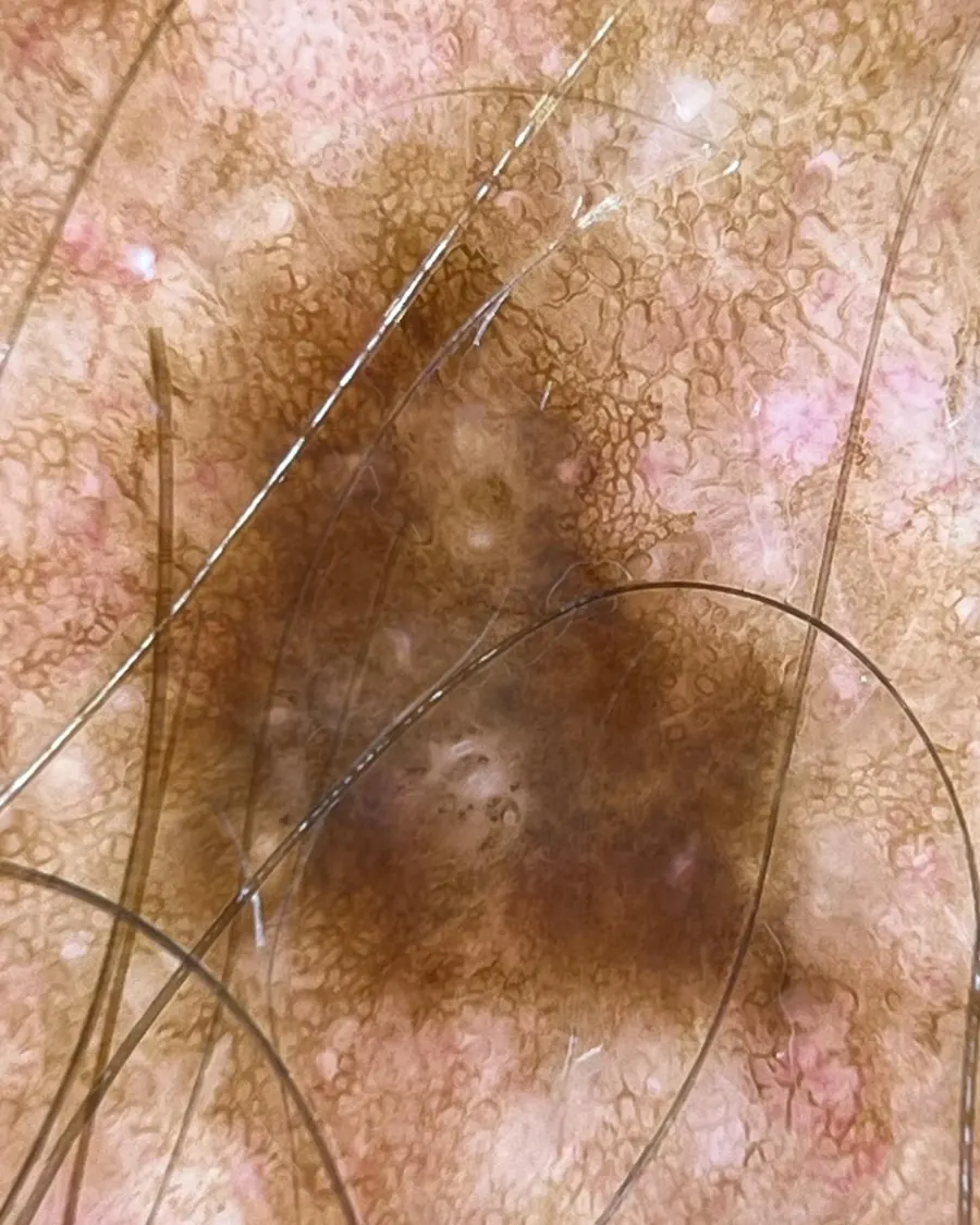
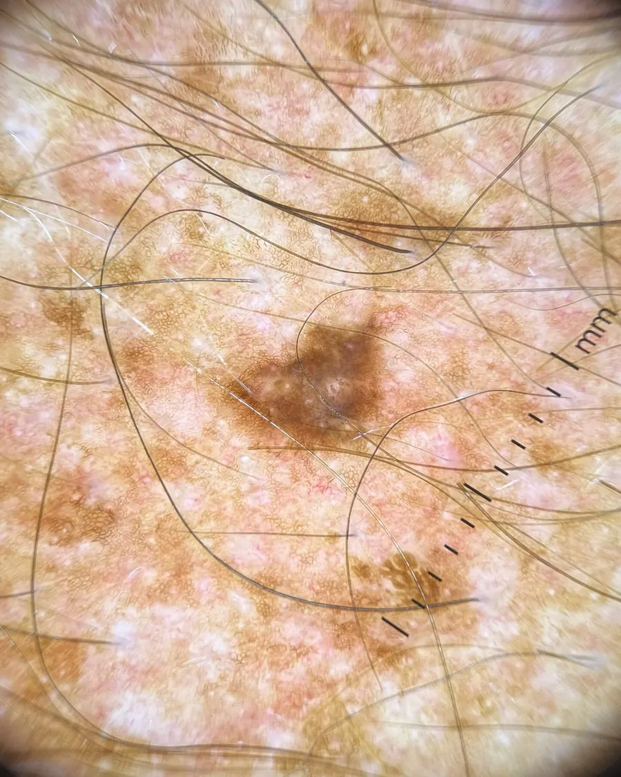
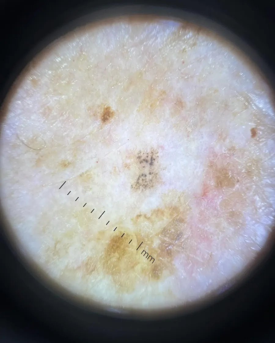
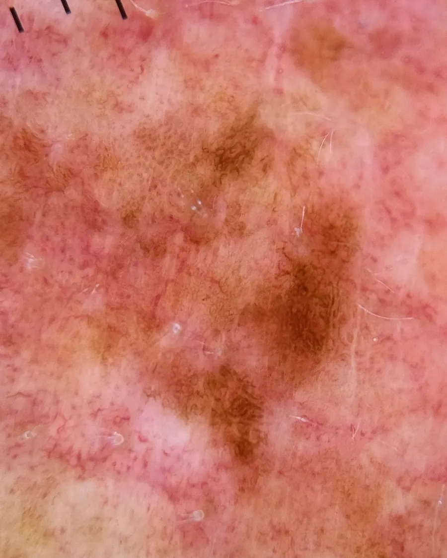
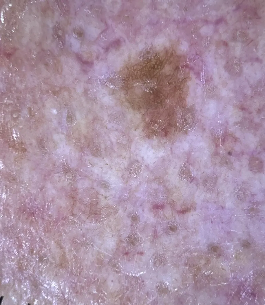
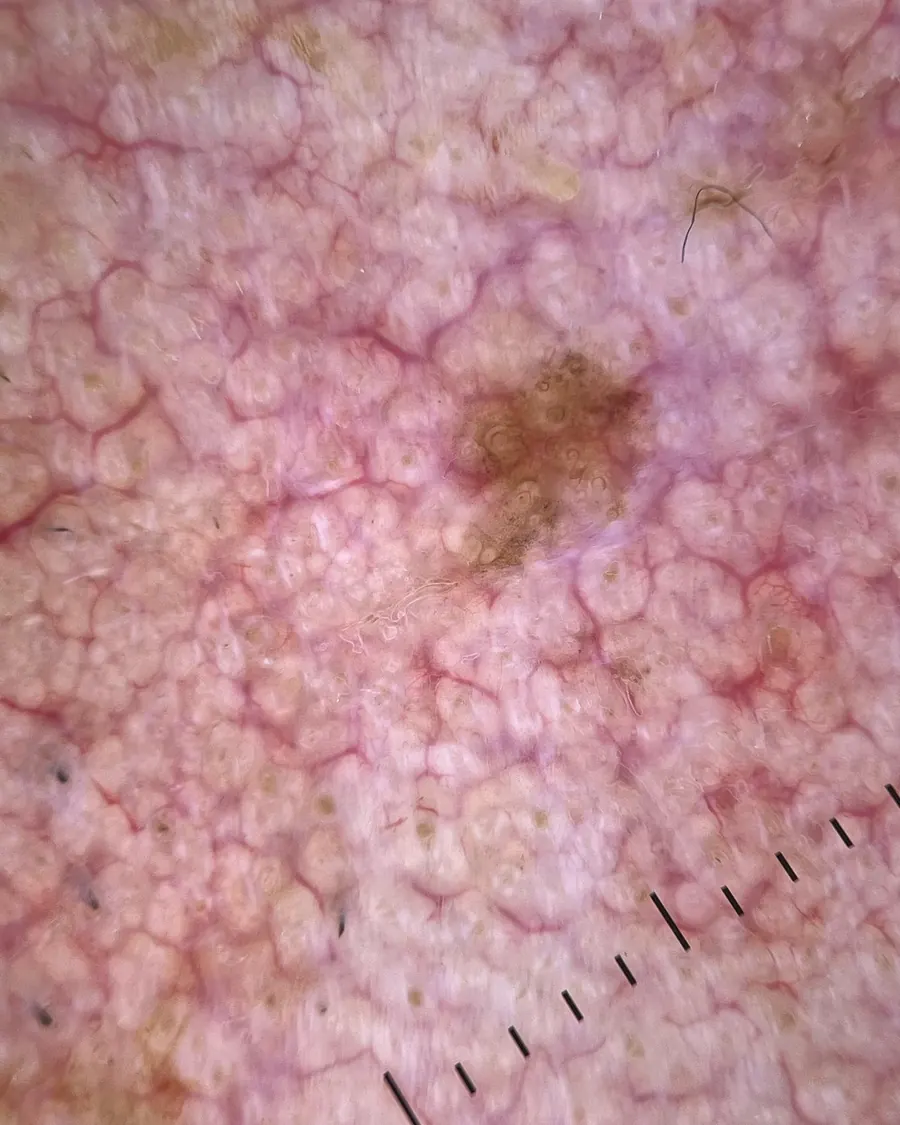
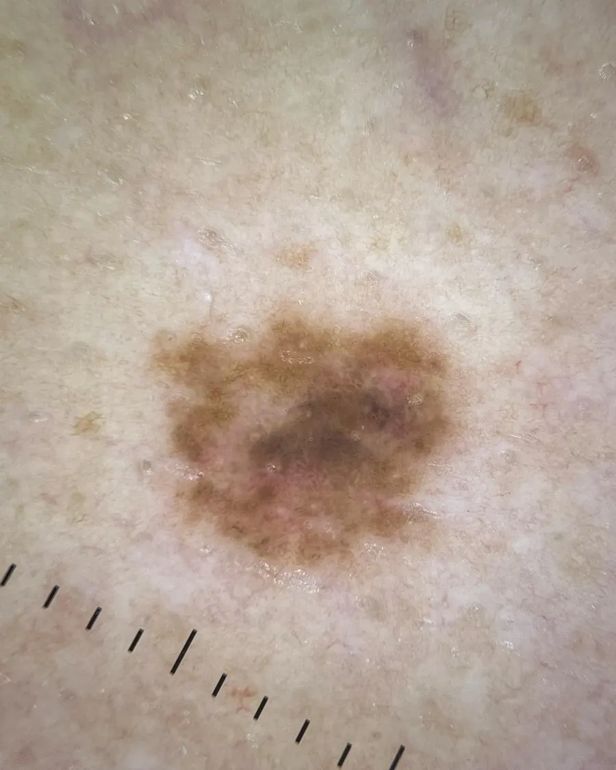
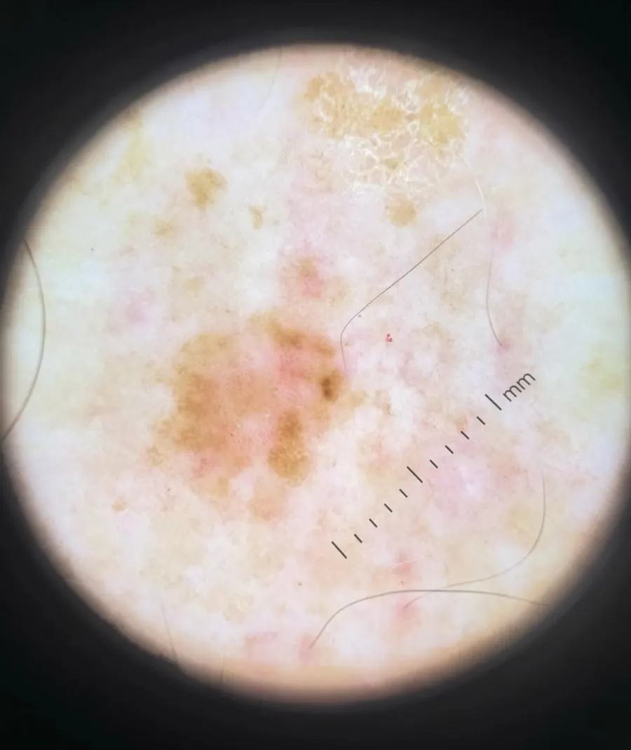
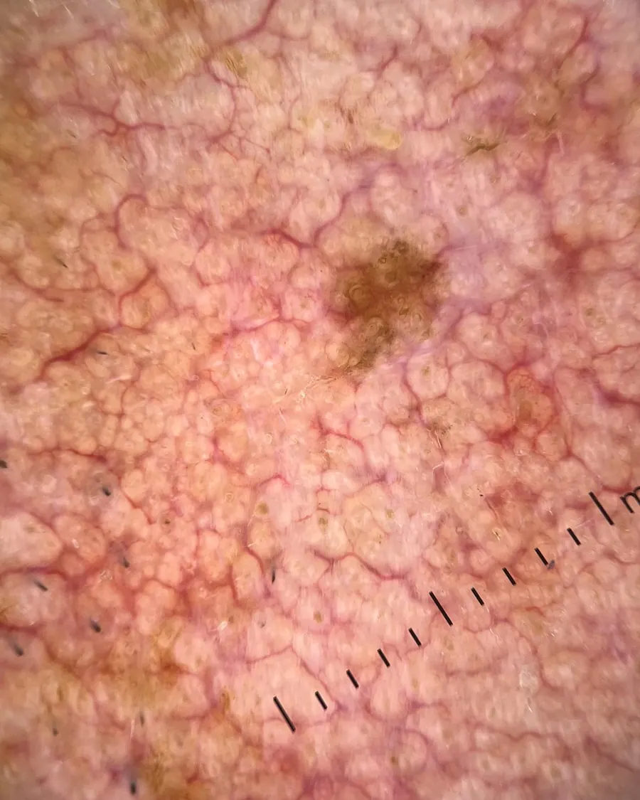
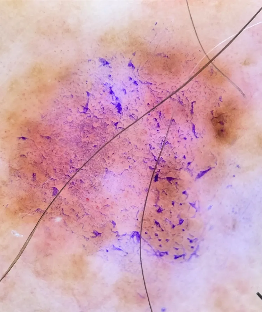
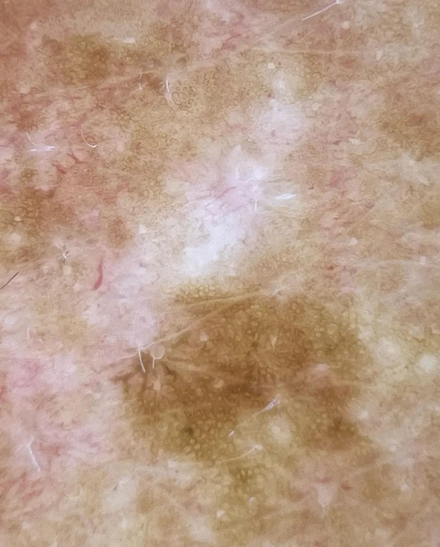
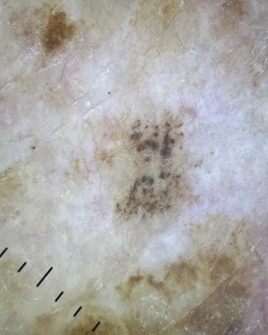
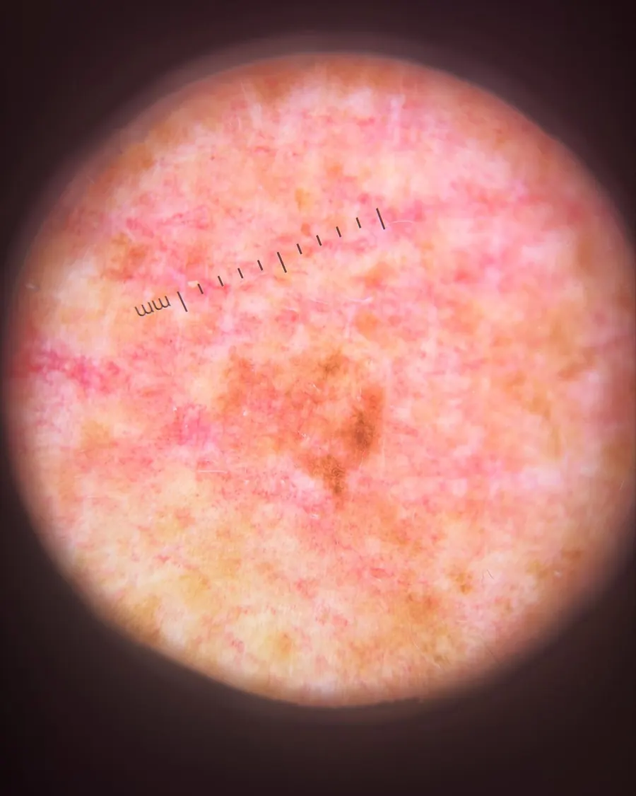
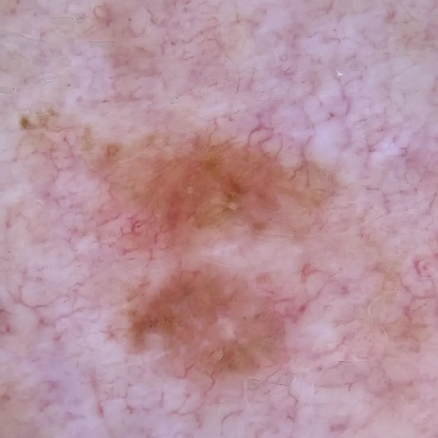
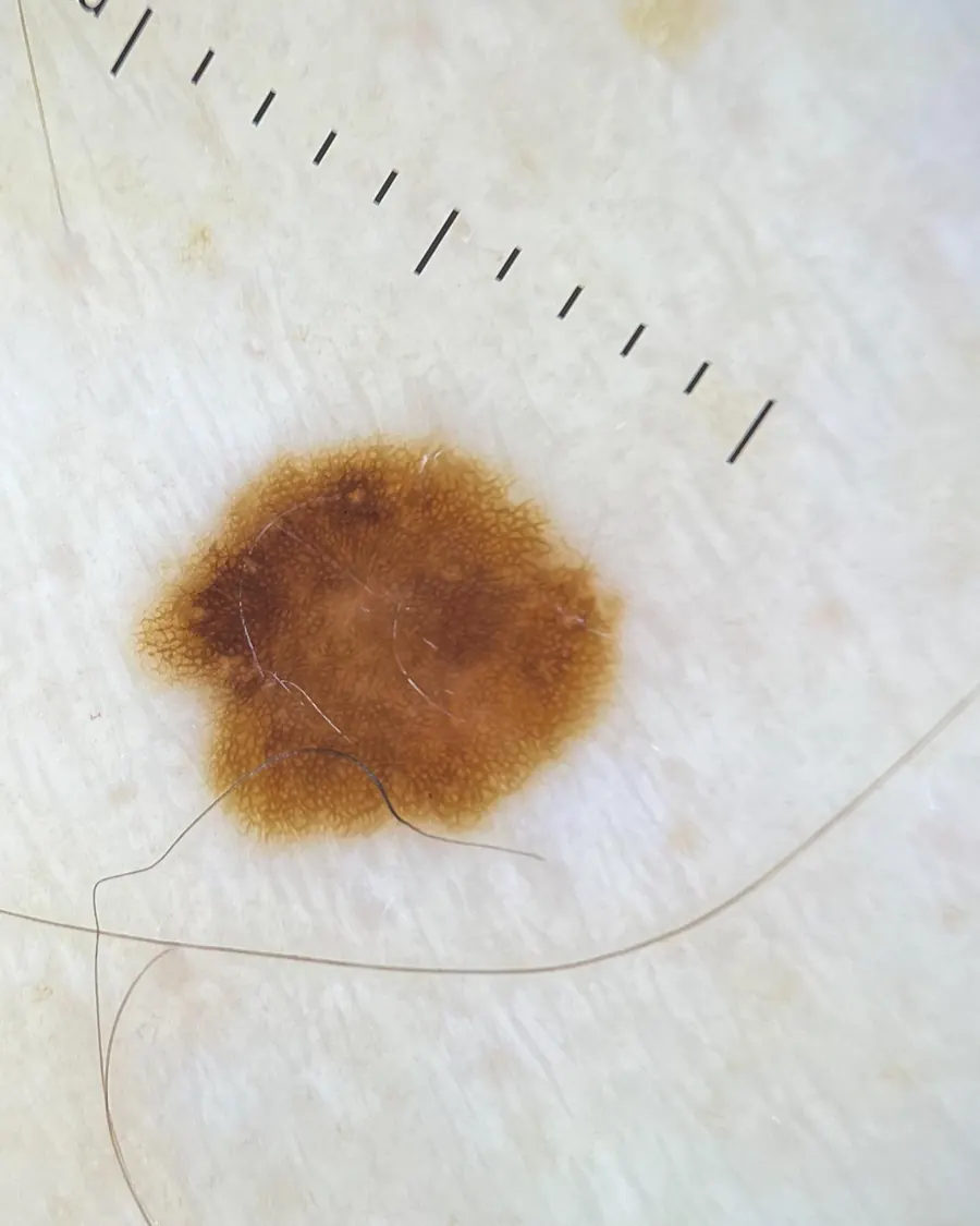
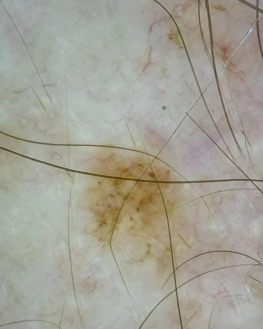
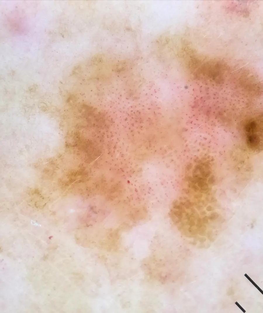
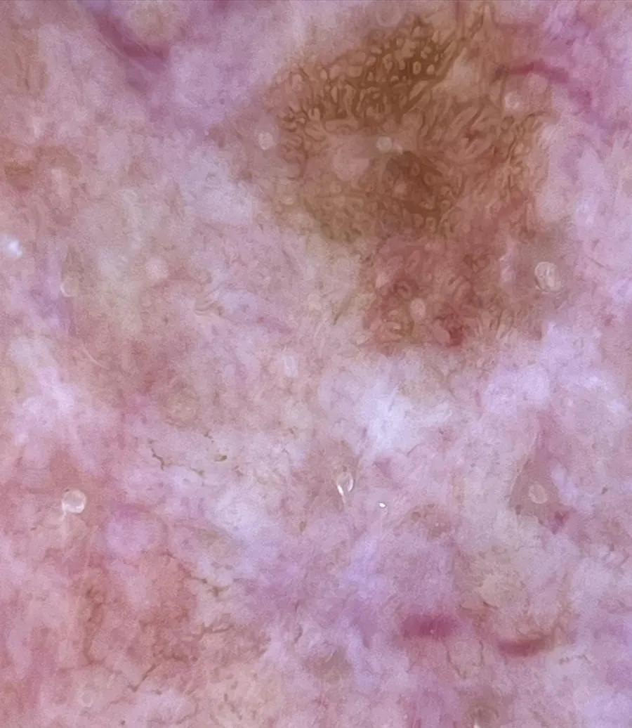
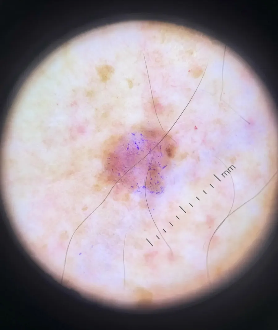
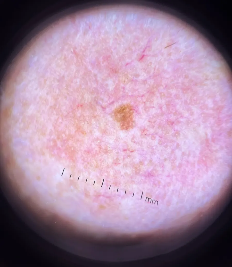
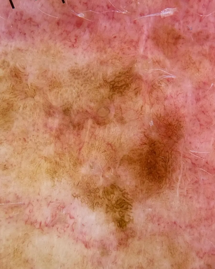
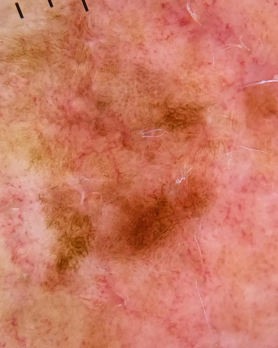
Many melanomas look normal. They can be round, symmetric, contain only one color, or be smaller than a freckle. An impossible task to spot with your eyes alone, because these melanomas are invisible to the naked eye.
A dermatoscope, applied to the skin, uses magnification with polarization to visualize subsurface structures inside the skin: vascular structures, network architecture, all important structures for rendering a mole as benign, or identifying it as concerning for melanoma. Most dermatologists own a dermatoscope. The gap is in how it’s used and how it’s taught.
02
Look for yourself.

03
Problem one: how it’s used.
Too often the dermatoscope only comes out for a spot that already looks suspicious. By then the exam has already failed. Most of the melanomas I identify only become concerning once they’re under the scope.
Using a dermatoscope to confirm a suspicion is a different exam from using it on every mole, every sunspot and every pink spot. I do not see more patients than other dermatologists, and I keep the number of skin exams in a day deliberately limited. What’s different is how I deploy my dermatoscope, and what I’ve learned to appreciate through it.
More than 95% of the melanomas I find during skin cancer screenings are spots my patients did not know were concerning.
Because most melanomas do not stand out. Finding them requires a defined strategy, with appropriate tactics: one that is deliberate, intentional and precise. That’s the standard I hold myself to.
04
people die of melanoma every year.4 A quarter of those deaths are in patients who had been deemed to be low risk.5
What if those “low-risk” tumors were found earlier still, as in situ disease, at stage zero? What if melanomas found at higher stages, III and IV, were found early?
Build the skin exam to find the melanoma that is invisible to the naked eye, and not just the melanoma that stands out.
05
Problem two: how it’s taught.
Dermoscopy is still often taught as an adjunct to aid in rendering a melanoma diagnosis, rather than the core skill needed to find melanoma. I finished residency in 2019. Dermatology residency runs four years, three of them in dermatology, and in that time I had two dedicated dermoscopy lectures of about an hour each. A discipline, necessary to identify most melanoma, passively instructed.
Understanding the pathology of skin biopsies, on the other hand, was taught extensively. We had weekly teaching sessions with a dermatopathologist, dedicated monthly dermatopathology rotations, and continuous exposure throughout residency. That emphasis was reinforced on our board examinations, as fifteen percent of the examination is dermatopathology.6
But this is the disconnect, from academics to real-world application. Most dermatologists do not review their pathology of biopsies after residency, as there is a fellowship to become a board-certified dermatopathologist. But most will conduct skin examinations to evaluate patients for skin cancer.
The skill that matters the most in the exam room to identify melanoma is treated as tertiary. In a 2017 survey, 38% of dermatology residents had received no dermoscopy training at all, and 91% wanted more.7
06
The numbers are public.
Arizona requires every melanoma to be reported to the state,8 and for four years running I’ve identified more melanomas than anyone else in Arizona (see the numbers). In 2023 that was 215, or 12.8× the state average.
My patients are much like any general dermatologist doing skin cancer screening in Arizona. The difference is in the exam, and my expertise of what was learned along the way.
07
What needs to change.
- 01
Teach dermoscopy at least as thoroughly as reading biopsy slides, and test for proficiency on board examination.
- 02
Require dermoscopy training for everyone who screens for skin cancer: physicians, physician assistants and nurse practitioners.
- 03
Structure every skin exam to deploy a dermatoscope on every mole, every sunspot and every pink spot.